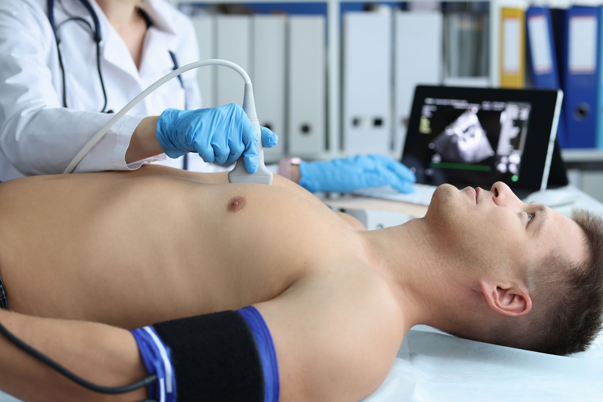
Echocardiogram
Introduction
An echocardiogram is usually also be referred to as a transthoracic echocardiogram (TTE), Doppler ultrasound of the heart, or surface echo. An echocardiogram is an ultrasound of the heart. During the procedure, sound waves create a “live” picture of the heart beating.
An echocardiogram is used to show a detailed moving picture of the heart. It is used to evaluate the functioning of the heart valves and chambers, assess heart pumping, and check for heart murmurs. An echocardiogram is commonly used to check for heart disease and evaluate the heart functioning of people that have had heart attacks.
Test Procedure
There is no special preparation prior to an echocardiogram. You will wear a hospital gown and disrobe from the waist up for the procedure. Conducting gel will be placed on your chest. A cardiologist or sonographer will place a transducer device on your chest. The device transmits sound waves to a monitor that produces moving pictures of your heart. In some cases, dye may be delivered through an IV to provide more contrast in the pictures. Your doctor will review the results with you. It is important to know that this procedure is different than a transesophageal echocardiogram (TEE) in which the ultrasound probe is passed through the mouth into the feeding tube to take images of the heart.

