Ultrasound - Diagnostic
Introduction
An ultrasound is an imaging test that allows doctors to view internal organs, tissues, and systems, and check developing babies during pregnancy. Ultrasound is noninvasive. An ultrasound uses sound waves to create pictures of the structures on a video monitor. Ultrasound does not use radiation like X-rays and there are no known risks associated with the procedure.
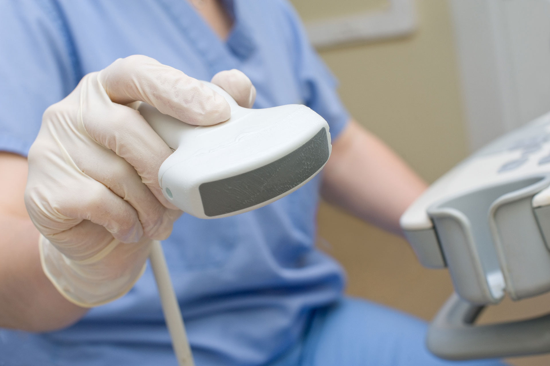
An ultrasound uses a small conduction device that is placed and moved on the skin. The conduction device transmits high-frequency sound wave information that is translated into pictures on a monitor. Particular images may be saved in a computer or printed out.
Preparation
Ultrasound may be performed in a doctor’s office, outpatient radiology center, or the radiology department of a hospital. Depending on the body area being assessed, you may need to wear a gown for the procedure.
In preparation for pelvic or obstetrical ultrasounds and kidney or renal ultrasounds you may need to drink fluids before the procedure and not urinate until after the procedure. A gallbladder, liver, abdominal, or aorta ultrasound may require fasting for eight hours. There is generally no special preparation for other types of ultrasound. You will receive specific preparation instructions when you schedule your appointment.
Procedure
You will lie on an examination table for the procedure. A conducting gel will be placed on your skin. Your doctor or radiology technician will gently place and move the conduction device across your skin over the area that is being imaged. An ultrasound creates little or no discomfort. When the test is complete, the conduction gel is wiped away.
If your doctor performs your ultrasound, your results may be discussed at the time of your test. A radiology technician may perform your test, but is not qualified to diagnose or discuss your condition or results with you. In this case, a radiologist or your doctor will review your results with you.

